Cervical Spine x-ray
Cervical Spine X-Ray (C-Spine X-Ray) is performed to investigate neck pain, cervical pain particularly following trauma or in cases of chronic neck pain with findings of upper limb weakness, numbness or tingling. A cervical spine X-ray is a safe and painless test that uses a small amount of radiation to take a picture of the bones in the back of the neck (cervical vertebrae).
Cervical Spine X-Ray (C-Spine X-Ray) is useful in identifying cervical vertebrae fractures (bone breaks), vertebral malalignment, dislocation and degenerative spine disease. A cervical spine X-ray can help find the cause of symptoms such as neck, shoulder, upper back, or arm pain, as well as tingling, numbness, or weakness in the arm or hand. It can detect fractures in the cervical vertebrae or dislocation of the joints between the vertebrae.
Although the Cervical Spine Pain X-Ray procedure may take up to 15 minutes, actual exposure to radiation is usually only a few seconds. The patient will be asked to bend the head forward as far as possible, and to extend the neck backwards as far as possible.
Cervical Spine X-Ray (C-Spine X-Ray) is performed by a radiographer in an X-Ray room. During the examination, an X-ray machine sends a beam of radiation through the neck, and an image is recorded on special film or a computer. This image includes the seven vertebrae in the neck area, the first vertebrae of the thoracic spine, and the disk spaces in between them.
The X-ray image is black and white. Dense body parts that block the passage of the X-ray beam through the body, such as the bones, appear white on the X-ray image. Hollow body parts, such as the airways, allow X-ray beams to pass through them and appear black.
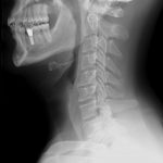
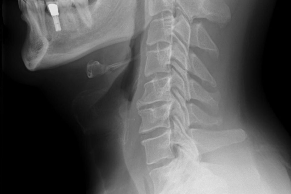
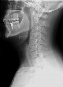
An X-ray technician takes the X-rays. Usually, three different pictures usually are taken of the cervical spine: one from the front (AP or anterior-posterior view), one from the side (lateral view), and another from the front through an open mouth (odontoid view). Occasionally, additional pictures like flexion and extension views of the cervical spine might be needed.
Cervical Spine X-Ray will be looked at by a radiologist (a doctor who is specially trained in reading and interpreting X-ray images). The radiologist will send a report to your doctoring cases of trauma, the Cervical Spine X-Ray is usually interpreted immediately by an emergency doctor such as an Emergency Physician, an Orthopedic Surgeon, or a General Surgeon involved in the care of the patient. Cervical Spine X-Ray Also Known As C-spine X-Ray, Cervical pain X-ray, C-spine series, AP cervical spine, Lateral cervical spine, Flexion/extension views, 5-series cervical spine, 7-series cervical spine.